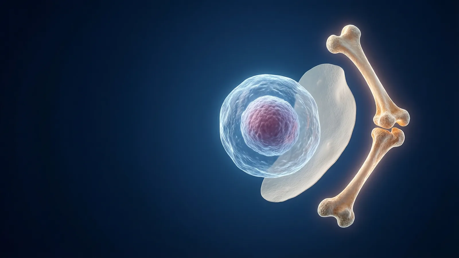
How the Human Body Is Organized: Cells, Tissues, Bones
See how cells build tissues, tissues build organs, and how the skeleton works as a living frame of bones, joints, cartilage and ligaments. With a live 3D lab.
Your body is not one machine but a stack of layers, each built from the one below. Trillions of tiny cells form a handful of tissues, tissues form organs, and organs team up into systems. Here is how those layers fit together, and why the skeleton is a much busier place than a museum display suggests.
Why study structure and function together?
Anatomy describes what the body is made of and where each part sits. Physiology asks how those parts behave while you are alive. The two questions are tied together: a thin, flat sheet of cells suits a surface where gases pass through, and a thick rope of fibres suits something that must pull.
Whenever you meet a new body part, ask two things. What is its shape? And what job does that shape make easy? The answers tend to match, and that habit makes a long list of names much easier to remember.
Level one: the cell
The cell is the smallest unit of life in the body. An adult is built from tens of trillions of them. Each cell is wrapped in a membrane that controls what goes in and out, and inside it are tiny working parts called organelles.
- The nucleus holds the instructions (DNA).
- Mitochondria release usable energy from fuel.
- Ribosomes assemble proteins.
Nearly every cell in your body carries the same set of instructions, yet a muscle cell looks and behaves nothing like a nerve cell. The difference is which instructions are switched on. A cell becomes specialised, and specialisation is what makes the next level possible.
Level two: four kinds of tissue
A tissue is a group of similar cells working as a team. One muscle cell can shorten only a tiny amount, but thousands lined up can move a limb. Every organ in the body is built from just four tissue types:
- Epithelial tissue covers surfaces and lines cavities, forming a barrier or a filter.
- Connective tissue supports, binds and fills. Bone, cartilage, fat and blood all count.
- Muscle tissue contracts to produce movement.
- Nervous tissue carries electrical signals.
Notice that bone and blood are both connective tissue, even though one is rock-hard and the other is liquid. What they share is that their cells sit in a matrix, the material between cells, and the matrix does much of the work.
Levels three and four: organs and systems
An organ combines two or more tissue types into one unit with a defined job. The stomach is a good example. Muscle churns the food, epithelium lines the inside and makes secretions, connective tissue holds the layers together, and nerves coordinate the whole thing.
Organs that cooperate on a larger task form an organ system: the digestive system, the circulatory system, the nervous system and so on. No system works alone. The skeleton protects the lungs, the lungs supply oxygen to the muscles that move the skeleton, and the nervous system directs all of it.
Keeping this teamwork steady is called homeostasis: holding conditions such as temperature and blood sugar within a narrow range even when the outside world changes.
The skeleton: a living frame
A typical adult skeleton has about 206 bones. They give the body its shape, hold it up against gravity, protect soft organs and provide rigid levers for muscles to pull on. Anatomists sort the bones into two groups:
- The axial skeleton: skull, spine and rib cage. It is built mostly for protection and support along the body's central line.
- The appendicular skeleton: the arms and legs plus the shoulder and hip girdles that attach them. It is built mostly for movement.
Bone is alive
Bone is a living connective tissue with its own blood supply. Specialised cells keep breaking down old bone and laying down new, which is called remodelling. This is how bone repairs itself and adjusts to the loads placed on it. Minerals give bone its hardness, and a flexible protein framework keeps it from being brittle.
Joints, cartilage and ligaments
A joint is any place where bones meet. Some joints, like the skull sutures, barely move. Others, like the shoulder, move in many directions. Smooth cartilage caps the bone ends and reduces friction, and tough ligaments link bone to bone and stop joints bending the wrong way.
Common misconceptions
- "Bones are dead and dry." Only in a museum. In a living person they are active tissue.
- "Each organ belongs to exactly one system." Many organs serve two. The pancreas, for instance, takes part in both digestion and hormone control.
- "Cells are all roughly the same." They share a basic plan but differ enormously in shape and job.
The 3D lab here is a simplified teaching model, not a medical reference, so use it to learn the ideas and the vocabulary rather than to judge anything about a real person's health.
How to explore this in the lab
Work from the smallest layer upward, then switch to the skeleton:
- The cell to see what a cell is made of.
- The four tissue types to learn the building materials of every organ.
- Organs and systems to watch tissues combine into working units.
- The skeleton for the overview of bones and their jobs.
- Bone as living tissue to see how bone renews itself.
- Joints to see how a rigid frame becomes a flexible body.
Rotate and zoom the model as you go. Seeing a structure from several sides fixes it in memory far better than reading its name.
- The body is built in layers: cells, tissues, organs, organ systems.
- Every organ is made from just four tissue types.
- Structure and function match, so shape is a clue to job.
- Bone is living, constantly remodelling connective tissue.
- Joints, cartilage and ligaments turn a rigid frame into a mobile one.
The cell
The smallest unit of life. About 6 minutes. Free, no account, runs in your browser.
More free lessons from this guide
Simulations on Novareka are teaching models, simplified on purpose. They show how an idea behaves; they are not a substitute for professional, medical or engineering advice.
Labs in this guide
 Live
Live Live
LiveKeep reading
All guides → Exam prep
 Explainer
 Explainer